 ALERT
ALERT ATTENTION ⚠️
In observance of a holiday, Agilent CrossLab/iLab Operations Software Support Help Desk will be closed during U.S. hours on Friday, June 19th, 2026. We will resume regular U.S. support hours on Monday, June 22nd, 2026. EU and APAC Support will remain open during this time. For urgent matters, please add "Urgent" to the ticket/email subject or press "1" when prompted to escalate a call on the iLab Support phone, and we will prioritize those requests first.
ALERT
The High Resolution Electron Microscopy Facility (HREMF) provides a resource to the scientific community at MD Anderson for high resolution imaging of cells, tissues, organs or polymers containing cancer agents. The facility is located at the Smith Research Building (South Campus) and houses a JEOL JEM-1010 transmission electron microscope (TEM), a JEOL JSM-5900 scanning electron microscope (SEM) equipped with a backscattered electron detector, a Bal-Tec Technotrade coating system, a Leica Ultramicrotome, a Leica Ultrostainer and other ancillary accessories needed to prepare samples for SEM and TEM. A technologist with histological training is available to assist researchers in defining their specific needs related to SEM and TEM. Both electron microscopes are equipped with digital cameras. The facility operates on a charge-back basis only for processing of samples and the number of microscope hours used to screen samples with technical assistance.
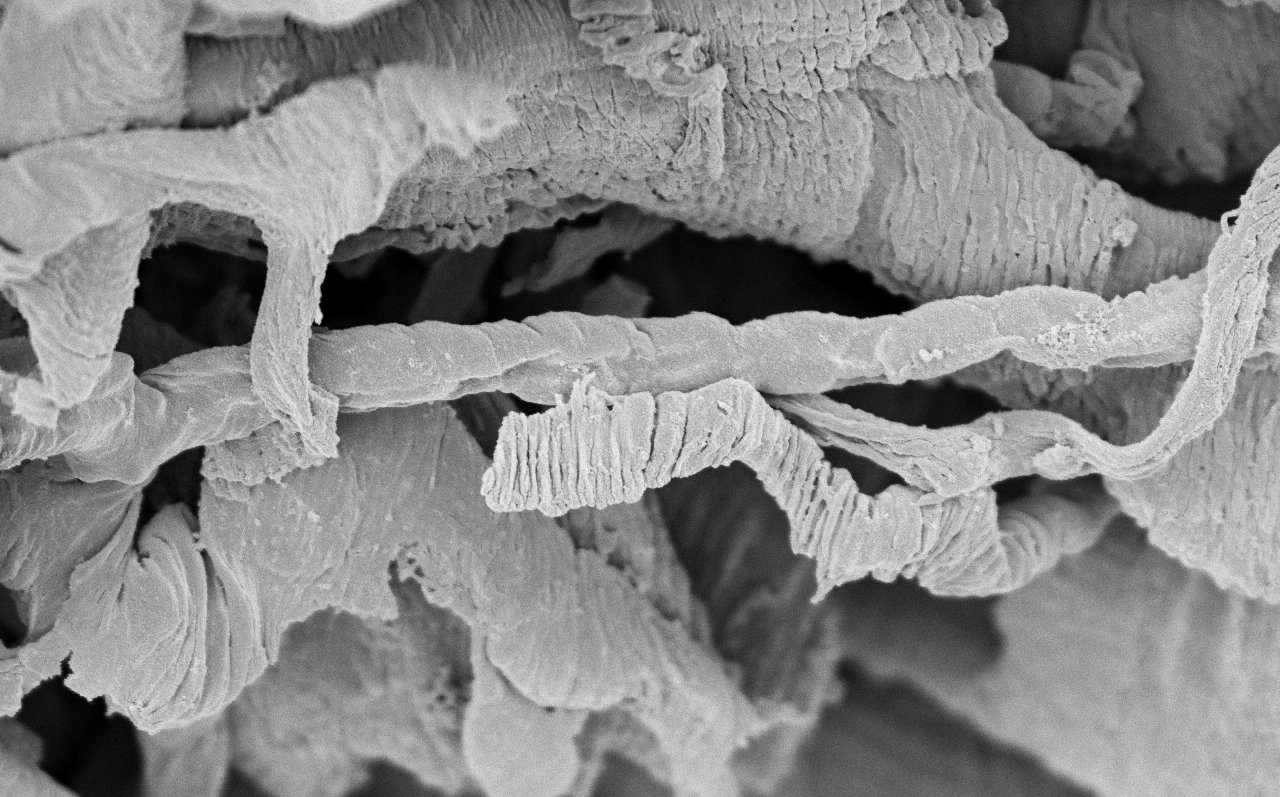
Scanning electron micrograph of the coronary microvasculature of a mouse that has been treated with a small molecule tyrosine kinase inhibitor of platelet-derived growth factor receptor beta. Image courtesy of Dr. Aarif Khakoo.
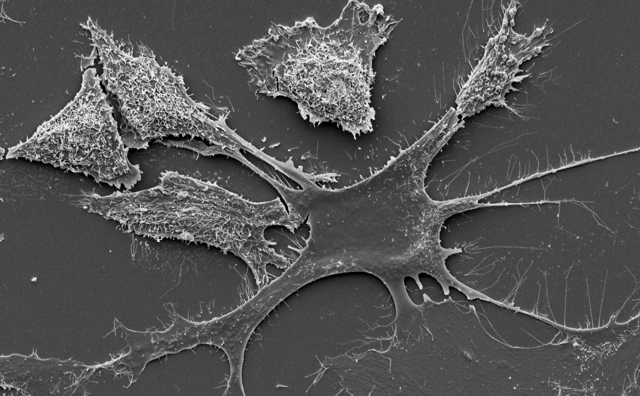
Scanning electron micrograph of breast cancer cells interacting with an astrocytes. Astrocytes upregulate survival proteins in cancer cells which protect those cells against chemotherapeutic agents such as temozolamide. Image courtesy of Dr. Isaiah Fidler.
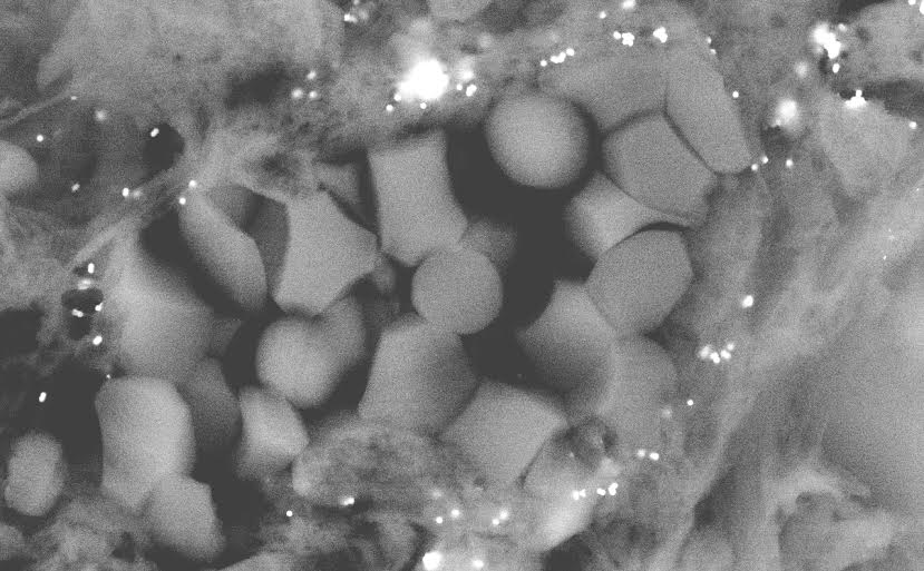
Scanning electron micrograph of gold nanoparticles localizing to the microcirculation of an experimental model of breast cancer. The blood vessel lumen contains several red blood cells and the nanoparticles are the small white structures.
Jian Hu, PhD, Core Director
Kenneth Dunner, Jr., A.S., B.S., Core Manager
|
Hours |
Location |
|
6:00am - 3:00pm Monday - Friday |
Smith Research Building |
| Name | Role | Phone | Location | |
|---|---|---|---|---|
| Kenneth Dunner, Jr., A.S., B.S. |
Core Manager
|
713-792-8109
|
kdunnerj@mdanderson.org
|
Smith Research Building, SRB1.688b
|
| Jian Hu, PhD |
Core Director
|
713-794-5238
|
jhu3@mdanderson.org
|
So Campus Research Bldg 3, 3SCR5.3612
|
| Services & Price List |
| Name | Description | Price |
|---|---|---|
| Cellular Ultrastructure Morphology |
Hourly Rate
For routine TEM imaging of cell and tissue structure and subcellular ultrastructure. |
Inquire |
| Exosome Characterization Studies |
Hourly Rate
Morphological, size, and protein assessment (immunogold labeling). Provides valuable information regarding sample purity and gives an overview of the level of contamination of the sample, for example with larger vesicles, such as microparticles, apoptotic bodies or cell debris. |
Inquire |
| Miscellaneous Charge |
Exact charge will be dependant upon the specifics of your request. |
Inquire |
| Monitoring Autophagy |
Hourly Rate
TEM analysis for detection of autophagy and quantification of autophagic accumulation. |
Inquire |
| Nanoparticles/nanostructures and Negative Staining |
Hourly Rate
For imaging small, 3-D nanoparticles/nanostructures adhering to grids. |
Inquire |
| Surface Morphology |
Hourly Rate
For routine SEM imaging of cell and tissue surface structure. |
Inquire |
| Unassisted Use | Inquire |